

What Are 3 Ways Enzymes Control Reactions
4.2: Control of Enzymatic Activeness
- Folio ID
- 7823
A printable version of this department is here: BiochemFFA_4_2.pdf. The entire textbook is bachelor for free from the authors at http://biochem.science.oregonstate.edu/content/biochemistry-free-and-easy
Regulation of enzyme activity
Autonomously from their ability to greatly speed the rates of chemical reactions in cells, enzymes have another holding that makes them valuable. This property is that their activity can be regulated, allowing them to be activated and inactivated, as necessary. This is tremendously of import in maintaining homeostasis, permitting cells to respond in controlled ways to changes in both internal and external conditions.
Inhibition of specific enzymes by drugs can also exist medically useful. Understanding the mechanisms that control enzyme activity is, therefore, of considerable importance.
Inhibition
We volition first discuss iv types of enzyme inhibition – competitive, not-competitive, uncompetitive, and suicide inhibition. Of these, the first three types are reversible. The last i, suicide inhibition, is not.
Competitive inhibition
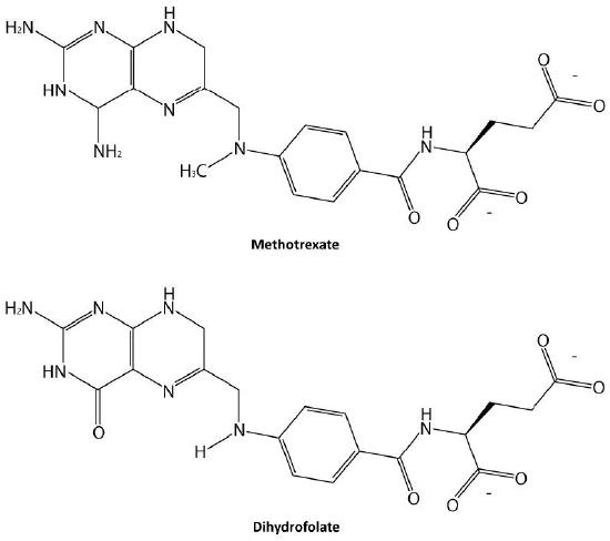
Probably the easiest type of enzyme inhibition to understand is competitive inhibition and it is the i virtually commonly exploited pharmaceutically. Molecules that are competitive inhibitors of enzymes resemble 1 of the normal substrates of an enzyme. An example is methotrexate, which resembles the folate substrate of the enzyme dihydrofolate reductase (DHFR). This enzyme commonly catalyzes the reduction of folate, an important reaction in the metabolism of nucleotides.
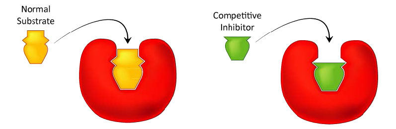
Figure four.33 - Competitive inhibitors resemble the normal substrate and compete for binding at the active site. Image past Aleia Kim
Inhibitor bounden
When the drug methotrexate is present, some of the DHFR enzyme binds to it, instead of to folate, and during the fourth dimension methotrexate is bound, the enzyme is inactive and unable to bind folate. Thus, the enzyme is inhibited. Notably, the bounden site on DHFR for methotrexate is the active site, the same identify that folate would normally demark. As a result, methotrexate 'competes' with folate for binding to the enzyme. The more methotrexate there is, the more effectively it competes with folate for the enzyme's active site. Conversely, the more folate there is, the less of an effect methotrexate has on the enzyme considering folate outcompetes information technology.
No effect on Vmax
How do we written report competitive inhibition? Information technology is typically done as follows. First, 1 performs a set up of V0 vs. [S] reactions without inhibitor (twenty or so tubes, with buffer and constant amounts of enzyme, varying amounts of substrate, equal reaction times). V0 vs. [S] is plotted (Effigy 4.35 ruddy line), every bit well as i/V0 vs. one/[S] (Figure iv.36 green line). Side by side, a second set of reactions is performed in the same way every bit before, except that a fixed amount of the methotrexate inhibitor is added to each tube. At low concentrations of substrate, the methotrexate competes for the enzyme effectively, simply at loftier concentrations of substrate, the inhibitor will have a much reduced effect, since the substrate outcompetes information technology, due to its college concentration (recall that the inhibitor is at fixed concentration).

Graphically, the results of these inhibitor experiments are shown in Figure iv.35 (blue line) and Figure 4.36 (orangish line). Notice that at loftier substrate concentrations, the competitive inhibitor has essentially no effect, causing the \(V_{max}\) for the enzyme to remain unchanged. To reiterate, this is due to the fact that at high substrate concentrations, the inhibitor doesn't compete well. Withal, at lower substrate concentrations, it does.

Increased \(K_m\)
In competitively inhibited reactions, the credible Km of the enzyme for the substrate increases (\(-1/K_m\) gets closer to zero - red line in Figure 4.36) when the inhibitor is present compared to when the inhibitor is absent, thus illustrating the better competition of the inhibitor at lower substrate concentrations. Information technology may not exist obvious why we call the inverse Km the apparent Km of the enzyme. The reason is that the inhibitor doesn't actually change the enzyme'due south affinity for the folate substrate. It but appears to do so. This is because of the style that competitive inhibition works. When the competitive inhibitor binds the enzyme, it is effectively 'taken out of action.' Inactive enzymes have NO affinity for substrate and no activity either. We tin't measure Km for an inactive enzyme.
The enzyme molecules that are not leap by methotrexate tin can, in fact, bind folate and are active. Methotrexate has no effect on them and their Km values are unchanged. Why then, does Km appear higher in the presence of a competitive inhibitor? The reason is that the competitive inhibitor is having a greater issue of reducing the amount of agile enzyme at lower concentrations of substrate than information technology does at college concentrations of substrate. When the corporeality of enzyme is reduced, 1 must have more substrate to supply the reduced amount of enzyme sufficiently to become to Vmax/two.
It is worth noting that in competitive inhibition, the percentage of inactive enzyme changes drastically over the range of [S] values used. To showtime, at low [Southward] values, the greatest percentage of the enzyme is inhibited. At high [S], no meaning percentage of enzyme is inhibited. This is not e'er the case, every bit we shall see in non-competitive inhibition.
Non-competitive inhibition
A second type of inhibition employs inhibitors that do non resemble the substrate and bind not to the active site, but rather to a separate site on the enzyme (Figure 4.37). The effect of binding a not-competitive inhibitor is significantly different from binding a competitive inhibitor because at that place is no contest. In the example of competitive inhibition, the effect of the inhibitor could be reduced and eventually overwhelmed with increasing amounts of substrate. This was considering increasing substrate made increasing percentages of the enzyme active. With non-competitive inhibition, increasing the amount of substrate has no issue on the percentage of enzyme that is active. Indeed, in non-competitive inhibition, the percentage of enzyme inhibited remains the same through all ranges of [S].

This means, then, that non-competitive inhibition effectively reduces the amount of enzyme past the aforementioned fixed amount in a typical experiment at every substrate concentration used The effect of this inhibition is shown in Figure four.38 & iv.39. Every bit you can see, \(V_{max}\) is reduced in non-competitive inhibition compared to uninhibited reactions.
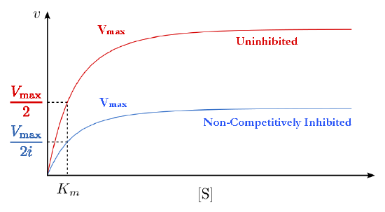
This makes sense if we think that Vmax is dependent on the amount of enzyme present. Reducing the amount of enzyme nowadays reduces \(V_{max}\). In competitive inhibition, this doesn't occur detectably, because at high substrate concentrations, there is essentially 100% of the enzyme agile and the \(V_{max}\) appears not to change. Additionally, Km for non-competitively inhibited reactions does non change from that of uninhibited reactions. This is because, as noted previously, i can only measure the \(K_m\) of active enzymes and \(K_m\) is a abiding for a given enzyme.

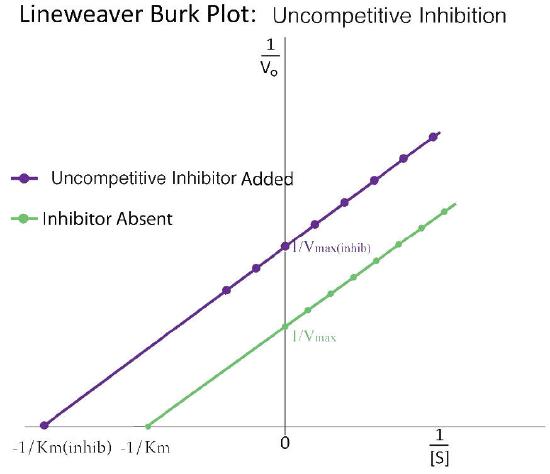
Uncompetitive inhibition
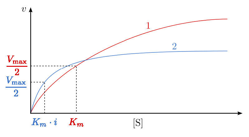
A third type of enzymatic inhibition is that of uncompetitive inhibition, which has the odd property of a reduced Vmax too as a reduced Km. The explanation for these seemingly odd results is rooted in the fact that the uncompetitive inhibitor binds only to the enzyme-substrate (ES) complex (Figure 4.forty). The inhibitor-leap circuitous forms mostly under concentrations of high substrate and the ES-I complex cannot release product while the inhibitor is spring, thus explaining the reduced \(V_{max}\).

The reduced Km is a scrap harder to anticipate. The reason is that the inhibitor-spring complex effectively reduces the concentration of the ES complex. By Le Chatelier's Principle, a shift occurs to form additional ES complex, resulting in less free enzyme and more enzyme in the forms ES and ESI (ES with inhibitor). Decreases in free enzyme correspond to an enzyme with greater affinity for its substrate. Thus, paradoxically, uncompetitive inhibition both decreases \(V_{max}\) and increases an enzyme's affinity for its substrate (\(K_m\) - Figures 4.41 & 4.42).
Suicide inhibition
In contrast to the kickoff iii types of inhibition, which involve reversible bounden of the inhibitor to the enzyme, suicide inhibition is irreversible, because the inhibitor becomes covalently bound to the enzyme during the inhibition. Suicide inhibition rather closely resembles competitive inhibition because the inhibitor by and large resembles the substrate and binds to the active site of the enzyme. The principal deviation is that the suicide inhibitor is chemically reactive in the active site and makes a bail with it that precludes its removal. Such a machinery is that employed past penicillin (Effigy iv.43), which covalently links to the bacterial enzyme, DD transpeptidase and stops it from performance. Since the normal office of the enzyme is to make a bond necessary for the peptidoglycan complex of the bacterial cell wall, the cell wall cannot properly course and bacteria cannot reproduce.

Control of enzymes
It is appropriate to talk at this point virtually mechanisms cells use to control enzymes. At that place are iv general methods that are employed:
- allosterism,
- covalent modification,
- access to substrate, and
- command of enzyme synthesis/breakup.
Some enzymes are controlled by more than one of these methods.
Allosterism
The term allosterism refers to the fact that the activity of certain enzymes can be afflicted by the binding of small molecules. Molecules causing allosteric effects come in two classifications. Ones that are substrates for the enzymes they affect are called homotropic effectors and those that are not substrates are called heterotropic effectors.
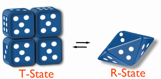
The homotropic effectors normally are activators of the enzymes they bind to and the results of their action tin can exist seen in the conversion of the hyperbolic bend typical of a V0 vs. [S] plot for an enzyme (Figure iv.18), beingness converted to a sigmoidal plot (Figure 4.44). This is due to the conversion of the enzyme from the T-state to the R-land on binding the substrate/homotropic effector.
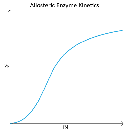
The V0 vs. [South] plot of allosteric enzyme reactions resembles the oxygen binding bend of hemoglobin (see Figure 2.83). Even though hemoglobin is not an enzyme and is thus not catalyzing a reaction, the similarity of the plots is non coincidental. In both cases, the binding of an external molecule is being measured – directly, in the hemoglobin plot, and indirectly by the V0 vs. [S] plot, since substrate binding is a factor in enzyme reaction velocity.
Allosteric inhibition
Allosterically, regulation of these enzymes works by inducing different physical states (shapes, as it were) that affect their ability to demark to substrate. When an enzyme is inhibited by binding an effector, it is converted to the T-state (T=tight), it has a reduced affinity for substrate and it is through this means that the reaction is slowed.
Allosteric activation
On the other paw, when an enzyme is activated by effector binding, information technology converts to the R-state (R=relaxed) and binds substrate much more readily. When no effector is present, the enzyme may exist in a mixture of T- and R-states.
Feedback inhibition
An interesting kind of allosteric control is exhibited by HMG-CoA reductase, which catalyzes an important reaction in the pathway leading to the synthesis of cholesterol. Binding of cholesterol to the enzyme reduces the enzyme'southward activity significantly. Cholesterol is non a substrate for the enzyme, and so it is therefore a heterotropic effector.
Notably, though, cholesterol is the stop-product of the pathway that HMG-CoA reductase catalyzes a reaction in. When enzymes are inhibited by an end-production of the pathway in which they participate, they are said to exhibit feedback inhibition.
Feedback inhibition always operates past allosterism and farther, provides important and efficient control of an entire pathway. By inhibiting an early enzyme in a pathway, the flow of materials (and ATP hydrolysis required for their processing) for the entire pathway is stopped or reduced, assuming there are not alternate supply methods.
Pathway control
In the cholesterol biosynthesis pathway, stopping this 1 enzyme has the event of shutting off (or at to the lowest degree slowing down) the entire pathway. This is significant because afterward catalysis by HMG-CoA reductase, there are over twenty further reactions necessary to make cholesterol, many of them requiring ATP energy. Shutting down ane reactions stops all of them. Another excellent instance of allosteric control and feedback inhibition is the enzyme ATCase, discussed below.
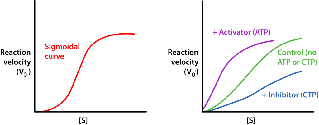
ATCase
Another interesting example of allosteric control and feedback inhibition is associated with the enzyme Aspartate Transcarbamoylase (ATCase). This enzyme, which catalyzes a step in the synthesis of pyrimidine nucleotides, has 12 subunits. These include half-dozen identical catalytic subunits and six identical regulatory subunits. The catalytic subunits bind to substrate and catalyze a reaction. The regulatory subunits bind to either ATP or CTP. If they demark to ATP, the enzyme subunits arrange themselves in the R-country.
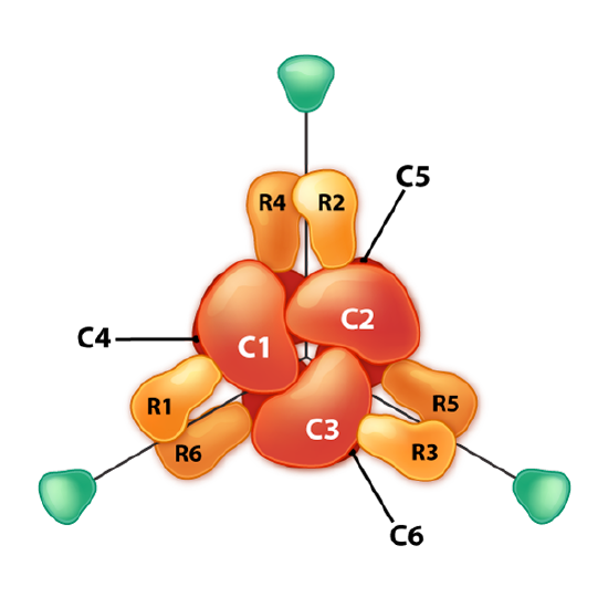
R-state
The R-state of ATCase allows the substrate to have easier access to the six agile sites and the reaction occurs more rapidly. For the aforementioned amount of substrate, an enzyme in the R-land will have a higher velocity than the aforementioned enzyme that is non in the R-land. Past dissimilarity, if the enzyme binds to CTP on i of its regulatory subunits, the subunits will adapt in the T-state and in this form, the substrate volition not have easy access to the active sites, resulting in a slower velocity for the aforementioned concentration of substrate compared to the R-state. ATCase is interesting in that it tin too flip into the R-state when one of the substrates (aspartate) binds to an active site within one of the catalytic subunits.
Aspartate has the outcome of activating the catalytic activeness of the enzyme by favoring the R-state. Thus, aspartate, which is a substrate of the enzyme is a homotropic effector and ATP and CTP, which are non substrates of the enzyme are heterotropic effectors of ATCase.
Allosteric models
In that location are three models commonly used to explain how allosterism regulates multi-subunit enzyme activity. They are known as
- the Monod-Wyman-Changeux (MWC) model (also known as the concerted model),
- the sequential model (also known equally KNF),
- and the morpheein model.
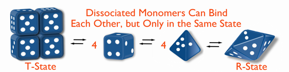
All models describe a Tense (T) country that is less catalytically agile and a Relaxed (R) state that is more than catalytically active. The models differ in how the states modify.
Sequential model
In the sequential model, bounden of an allosteric effector by one subunit causes it to change from T to R state (or vice versa) and that change makes information technology easier for adjacent subunits to similarly change state. With this model , there is a crusade/effect human relationship between binding of an effector by one subunit and change of state by an side by side subunit.
In hemoglobin, for example, binding of one oxygen past one unit of the circuitous may induce that unit of measurement to flip to the R-state and, through interactions with other subunits, cause them to favor adopting the R configuration earlier they bind to oxygen. In this way, binding of i subunit favors bounden of others and cooperativity tin exist explained by the alter in binding affinity as oxygen concentration changes.

MWC model
The MWC model is less intuitive. In it, the entire complex changes state from T to R (or vice versa) independently of the binding of effectors. Flipping betwixt T-states and R-states is postulated to be in an equilibrium of states in the absenteeism of effector (for instance, a l to 1 ratio of T/R. This ratio is referred to equally L, so L = T/R). Binding of effector past the enzyme complex has the tendency of "locking" the circuitous in a country. Bounden of inhibitors volition increase the ratio of T/R whereas binding of activators volition increase R and thus decrease the ration of T/R.
Morpheein model
The morpheein model is similar to the MWC model, but with an added step of dissociation of the subunits. The MWC model proposes that flipping between R and T states occurs past the complex as a whole and occurs on all units simultaneously. The morpheein model instead proposes that the multi-subunit enzyme breaks down to individual units which can then flip in construction and re-form the complex. In the morpheein model, only identically shaped units (all R, for example) tin can come together in the circuitous, thus explaining the "all-R-" or "all-T-" state plant in the MWC model.
A large number of enzymes, including prominent ones like citrate synthase, acetyl-CoA carboxylase, glutamate dehydrogenase, ribonucleotide reductase and lactate dehydrogenase take behavior consequent with the morpheein model.
Covalent control of enzymes
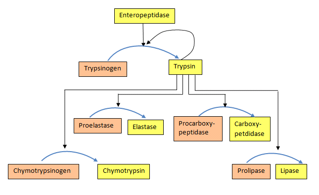
Some enzymes are synthesized in a completely inactive course and their activation requires covalent bonds in them to exist cleaved. Such inactive forms of enzymes are called zymogens. Examples include the proteins involved in claret clotting and proteolytic enzymes of the digestive system, such equally trypsin, chymotrypsin, pepsin, and others.
Synthesizing some enzymes in an inactive form makes very expert sense when an enzyme's activeness might exist harmful to the tissue where information technology is being fabricated. For example, the painful status known every bit pancreatitis arises when digestive enzymes made in the pancreas are activated besides soon and stop up attacking the pancreas.
Cascades
For both the claret clotting enzymes and the digestive enzymes, the zymogens are activated in a protease cascade. This occurs when activation of 1 enzyme activates others in a sort of chain reaction. In such a scheme the first enzyme activated proteolytically cleaves the 2d zymogen, causing information technology to be activated, which in plough activates a third and this may proceed through several levels of enzymatic action (Figure 4.50).
The reward of cascades is that they allow a large corporeality of zymogens to become activated adequately quickly, since in that location is an amplification of the bespeak at each level of catalysis.
Zymogens are also abundant in claret. Blood clotting involves polymerization of a protein known every bit fibrin. Since random formation of fibrin is extremely hazardous because it can block the menstruation of claret, potentially causing heart set on/stroke, the trunk synthesizes fibrin equally a zymogen (fibrinogen) and its activation results from a "pour" of activations of proteases that arise when a betoken is received from a wound. Similarly, the enzyme catalyzing removal of fibrin clots (plasmin) is also synthesized as a zymogen (plasminogen), since random clot removal would besides be chancy (see below likewise).
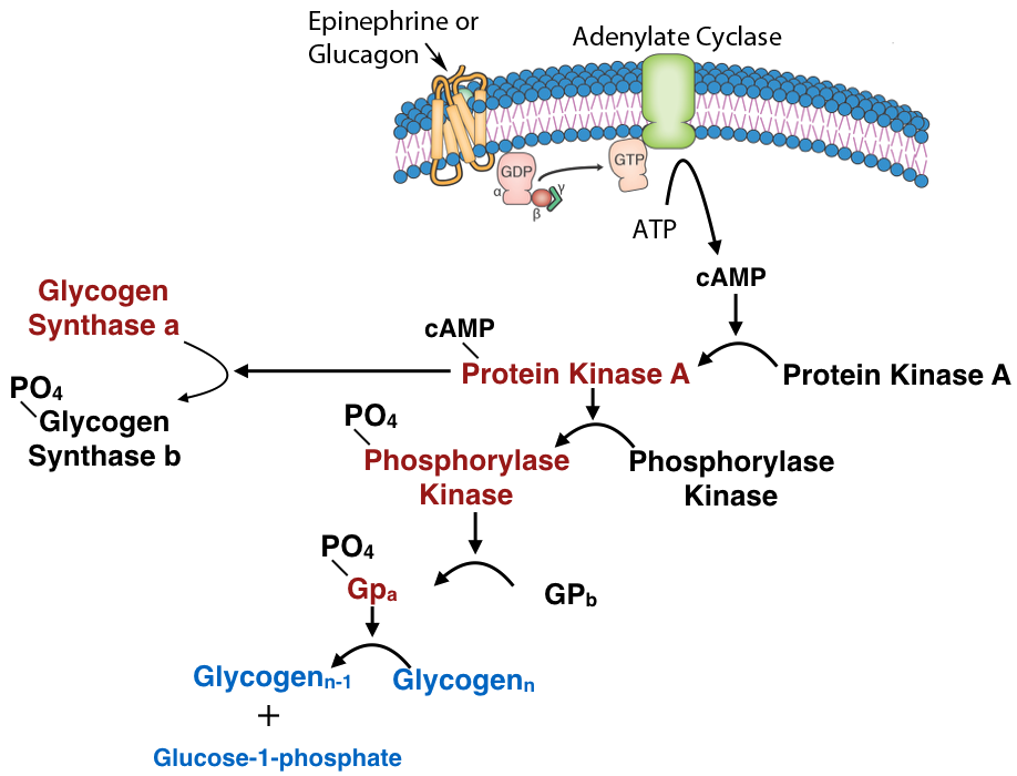
Phosphorylation/dephosphorylation
Another common mechanism for control of enzyme activity by covalent modification is phosphorylation. The phosphorylation of enzymes (on the side chains of serine, threonine or tyrosine residues) is carried out by protein kinases. Enzymes activated by phosphorylation can be regulated by the addition of phosphate groups by kinases or their removal by phosphatases. Thus, this type of covalent modification is readily reversible, in dissimilarity to proteolytic cleavage.
Reduction/oxidation
An interesting covalent control of enzymes using reduction/oxidation is exhibited in photosynthetic plants. In the light phase of photosynthesis, electrons are excited past light and flow through carriers to NADP+, forming NADPH. Thus, in the light, the NADPH concentration is high. When NADPH concentration is high, the concentration of reduced ferredoxin (a molecule donating electrons to NADP+) is likewise high.
Reduced ferredoxin can transfer electrons to thioredoxin, reducing it. Reduced thioredoxin can, in plough, transfer electrons to proteins to reduce their disulfide bonds. Four enzymes related to the Calvin wheel can receive electrons from thioredoxin and become activated, as a result.
These include sedoheptulose 1,7-bisphosphatase, ribulose-five-phosphate kinase, fructose 1,6-bisphosphatase, and glyceraldehyde three-phosphate dehydrogenase. Thus, in the calorie-free, electrons menstruation, causing NADPH to accumulate and ferredoxin to push electrons in the direction of these enzymes to a higher place, activating them and favoring the Calvin cycle. In the dark, the concentration of reduced NADPH, reduced ferredoxin, and reduced thioredoxin fall, resulting in loss of electrons by the Calvin bike enzymes (oxidations that re-form disulfide bonds) and the Calvin wheel inactivates.
Other enzyme control mechanisms
Other means of controlling enzymes chronicle to access to substrate (substrate-level control) and control of enzyme synthesis. Hexokinase is an enzyme that is largely regulated by availability of its substrate, glucose. When glucose concentration is low, the product of the enzyme's catalysis, glucose-vi-phosphate, inhibits the enzyme's function.
Regulation of enzymes past decision-making their synthesis is covered after in the book in the discussion relating to control of gene expression.

What Are 3 Ways Enzymes Control Reactions,
Source: https://bio.libretexts.org/Bookshelves/Biochemistry/Book%3A_Biochemistry_Free_For_All_(Ahern_Rajagopal_and_Tan)/04%3A_Catalysis/4.02%3A_Control_of_Enzymatic_Activity
Posted by: yearwoodlifeare.blogspot.com

0 Response to "What Are 3 Ways Enzymes Control Reactions"
Post a Comment